. 2023 Jun 15;24(6):510–523. doi: 10.1631/jzus.B2200447

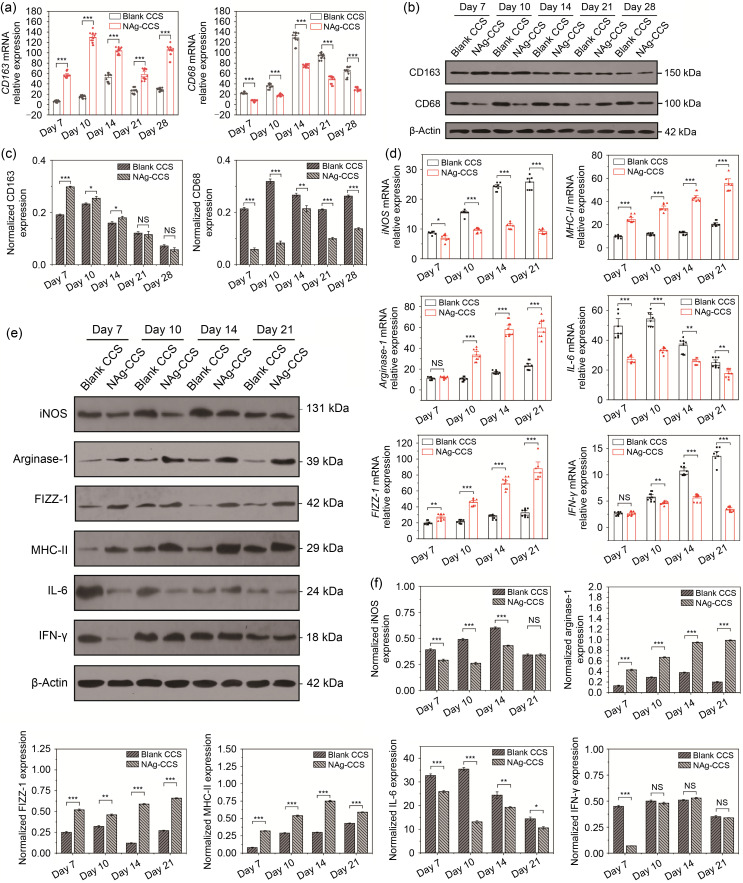
Fig. 6. Inflammation and macrophage infiltration after scaffold implantation in miniature pigs. (a) Relative mRNA expression of CD163 (left panel) and CD68 (right panel) was detected by qRT-PCR. (b) Representative images showing results for western blotting of CD163 and CD68. (c) Relative protein expression of CD163 (left panel) and CD68 (right panel). (d) Relative mRNA expression of iNOS, arginase-1, FIZZ-1, MHC-II, IL-6, and IFN-γ. (e) Representative images showing the western blotting results of iNOS, arginase-1, FIZZ-1, MHC-II, IL-6, and IFN-γ. (f) Relative protein expression of iNOS, arginase-1, FIZZ-1, MHC-II, IL-6, and IFN-γ. Data are expressed as mean±standard deviation (SD) ( n=3). * P<0.05, ** P<0.01, *** P<0.001; NS: not significant. NAg: nanosilver; CCS: collagen-chitosan scaffold; mRNA: messenger RNA; CD: cluster of differentiation; qRT-PCR: quantitative real-time polymerase chain reaction; iNOS: inducible nitric oxide synthase; FIZZ-1: resistin-like molecule alpha 1; MHC-II: major histocompatibility complex-II; IL-6: interleukin-6; IFN-γ: interferon-γ.

Fig. 6