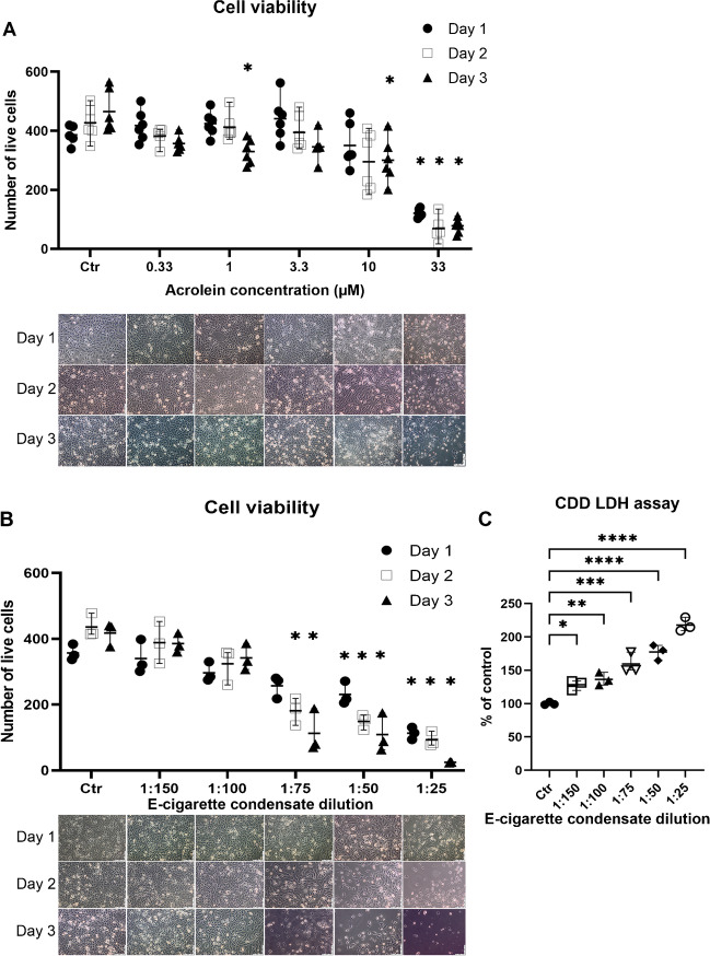
Fig. 2.
Effects of acrolein and E-cigarette condensate on EA.hy 926 cells. EA.hy 926 cells were incubated for 3 days with solvent (1% DMSO, named control) or A different concentrations of acrolein in DMSO (0.33, 1, 3.3, 10, 33 µM) or B E-cigarette condensate (1:150, 1:100, 1:75, 1:50, 1:25). Pictures were taken each day to observe the decrease in cell viability. The viable cells were counted from the pictures as the cells with a visible nucleus. The data are shown as mean ± range from n = 3–6 independent experiments. By recommendation of the statistical package from the Prism 9.0 software, a mixed-effects model (due to missing matched values) was used for data in panel A, whereas conventional 2-way ANOVA was used for data in panel B. Significance is indicated when p < 0.05 between the treated and control groups. C Cell death assay (lactate dehydrogenase [LDH]-based) was performed on the collected cell medium after the 3-day exposure to confirm the manual cell counts from images presented in panels A and B. The data are shown as mean ± range from n = 3 independent experiments. Conventional 1-way ANOVA analysis was performed. Significance is indicated as *, **, *** and **** when p < 0.05, p < 0.01, p < 0.001 and p < 0.0001 respectively

