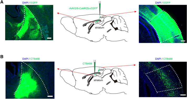
Fig. 5.
Neuronal projection from the AAF to the striatum. A Neuronal projection from the AAF to the striatum was revealed using EGFP. Middle, EGFP diagram tracing from the AAF to the striatum. Left, EGFP in the striatum. Right, The virus is microinjected into the AAF. Scale bars, 200 μm. B Neuronal projection from the striatum to the AAF revealed using CTB488. Middle, CTB488 diagram tracing from the striatum to the AAF. Left, CTB488 is microinjected into the striatum. Right, CTB488 signals in the AAF retrogradely filled from the striatum. Scale bars, 200 μm.

