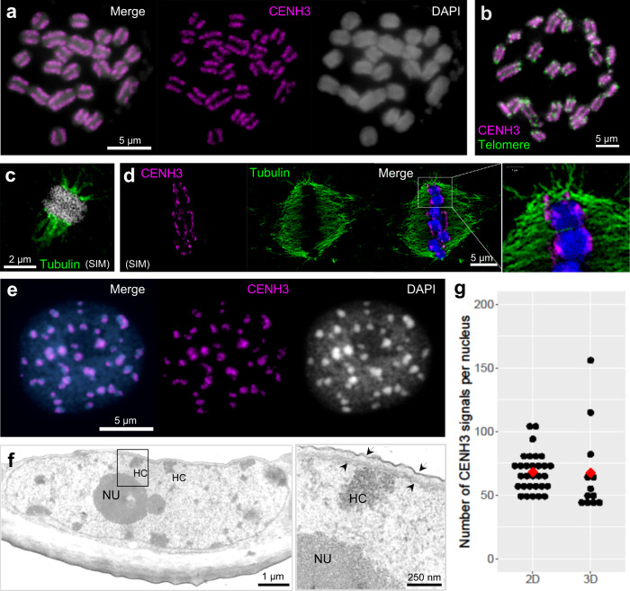
Fig. 1. C. japonica centromeres are distributed along entire mitotic chromosomes and form nuclear chromocenters.
a Condensed metaphase chromosomes show line-like CENH3 immuno-signals on the poleward surface of each chromatid, b from telomere to telomere. c Microtubules attach to the poleward surface of both chromatids. d Localization of CENH3 and tubulin sites. The enlargement shows the colocalization between CENH3 and microtubules. e CENH3 signals cluster in chromocenters of the interphase nucleus. c, d were taken by super-resolution microscopy (SIM). Chromosomes and nuclei were counterstained with DAPI. f Transmission electron micrograph of a C. japonica interphase nucleus. Electron-dense heterochromatic chromocenters (HC) are often located in the proximity of the double-layered nuclear membrane (further enlarged insert, arrows). NU, nucleolus. g The number of CENH3 signal clusters per interphase nucleus counted in 2D (n = 30) and 3D (n = 12) image stacks. Red dots show the average number. a–f At least two independent experiments were carried out to confirm the reproducibility of the labeling patterns. g Source data are provided as a Source Data file.

