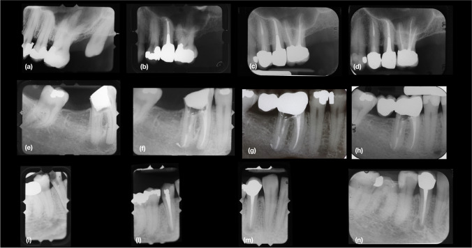
Fig. 1 .
(a), Tooth 2.5. pre-op periapical radiograph, year 1979. (b), Periapical radiograph after endodontic and restorative treatment with cast metal post and crown, year 1979. (c), Post-op periapical radiograph follow-up, year 2004. (d), Post-op periapical radiograph demonstrating success 37 years after primary endodontic treatment , year 2016. (e) Tooth 4.6. pre-op periapical radiograph, year 1982. (f), Periapical radiograph after endodontic treatment, year 1982. (g), Tooth 4.6. post-op periapical radiograph with restorative treatment with a fiber post and radiolucent composite material and a crown, follow-up year 2000. (h), Tooth 4.6. post-op periapical radiograph demonstrating success after 34 years follow-up, year 2016. (i), Tooth 4.4. pre-op periapical radiograph, year 1979. (l), Periapical radiograph after endodontic treatment, year 1979. (m), Post-op periapical radiograph follow-up with restorative treatment, year 2004. (n), Post-op periapical radiograph demonstrating survival in painless function 37 years after primary endodontic treatment, year 2016.

