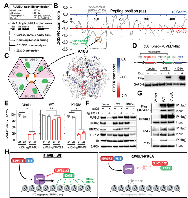
Figure 5.

Lysine 108 in RUVBL1 is required for the interaction between RUVBL1 and MYC. A) Schematic outline of RUVBL1 high‐density CRISPR gene body scan in A673‐Cas9 cells. B) 2D annotation of RUVBL1 CRISPR scan. The gray line indicates the smoothened model of the CRISPR scan score derived from 194 sgRNAs (dots) targeting the coding exons of RUVBL1 (n = 3 replicates). The median CRISPR scan scores of the positive control (red line; defined as −1.0) and negative control (blue line; defined as 0.0) sgRNAs are highlighted. C) 3D annotation RUVBL1 CRISPR scan score relative to a cryo‐EM structural model of a hexamer consists of three RUVBL1 and three RUVBL2 proteins (PDB ID: 5OAF). D) Western blot showing doxycycline (DOX)‐induced expression of flag‐tagged WT‐ and K108A‐RUVBL1 in A673‐dCas9‐KRAB cells. E) Effect of WT‐ and K108A‐RUVBL1 expression on the growth competition assay of A673‐dCas9‐KRAB cells transduced with sgiCtrl and sgiRUVBL1 (n = 3 for each group). F) Western blot of RUVBL1, H4K8ac, H4K12ac, EEF1A1, histone H4, and GAPDH in WT‐ and K108A‐RUVBL1 expressing A673‐dCas9‐KRAB cells transduced with sgiCtrl and sgiRUVBL1. G) Co‐IP of WT‐ and K108A‐RUVBL1 (flag‐tagged) with RUVBL2, KAT5, and MYC in HEK293 cells. H) Model of RUVBL1 supporting MYC chromatin binding and target gene expression. Data are represented as mean ± SEM. *P < 0.01 compared to sgCtrl by two‐sided Student's t‐test. Source data are available for this figure: SourceData F5 B.
