
Figure 1.
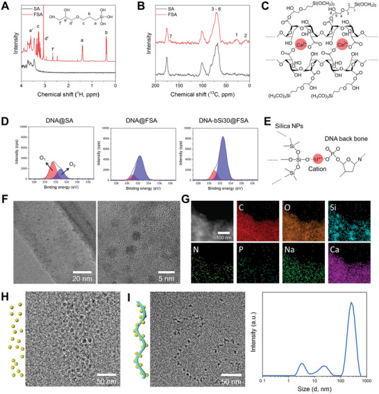
Results of A) liquid‐state 1H NMR and B) solid‐state 13C NMR of the pristine alginate and prepared functionalized alginate (FSA) hydrogel. C) Chemical structure of the prepared hydrogel inks comprising ionically crosslinked FSA based on NMR results. D) XPS results (O 1s) of DNA@SA, DNA@FSA, and DNA‐bSi30@FSA, and E) schematic diagram of DNA‐conjugated silica NPs. F) Representative TEM images of synthesized silica NPs in DNA@FSA ink at different magnifications. G) STEM and elemental mapping images of DNA@FSA ink. H) Silica NPs present in Si@FSA ink observed with cryo‐TEM. I) Biomineralized silica NPs on DNA with a ribbon‐like structure present in DNA‐bSi@FSA inks and their size distribution.