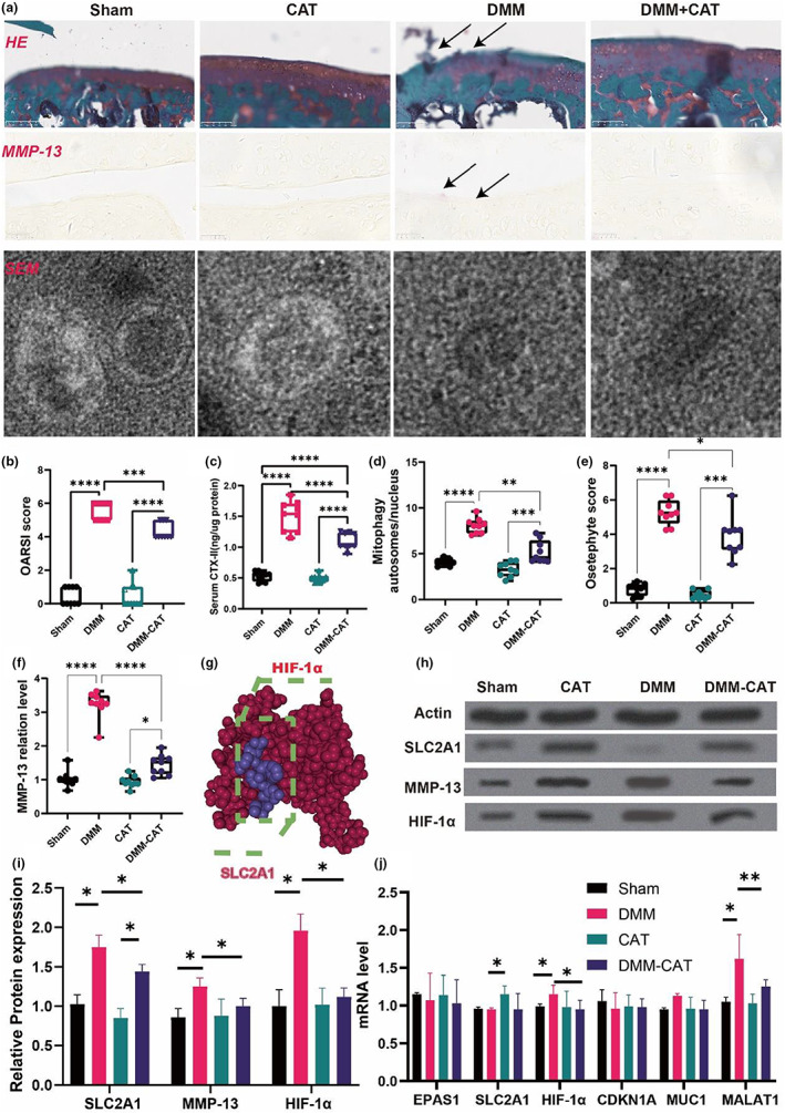
FIGURE 6.

CAT regulated the SLC2A1, MMP‐13, HIF‐1α expression and improved the development of OA mice. (a) Representative diagram of Safranin‐O/Fast Green staining, immunofluorescence, scanning electron microscope. (b) OARSI score. (c) serum CTX‐II. (d) Mitophagy autosomes/nucleus. (e) Osteophyte score. (f) MMP‐13 protein level. (g) 3D binding structure of HIF1 and SCL2A1 determined via molecular modeling and docking studies. (h) Western blot. (i) Relative protein expression. (j) mRNA level. All data are from n = 9 independent experiments. OA, osteoarthritis; CAT, Capsiate; AUC, Area Under The Curve; GSH, glutathione; GSH/GSSH, glutathione/oxidized glutathione; DXA, dual energy x‐ray absorptiometry; BV/TV, bone volume over total volume; Tb.Th, trabecular thickness; Tb.N, trabecular number; Tb.Sp, trabecular spacing; OARSI, Osteoarthritis Research Society International. MMP‐13, matrix metalloproteinase‐13; DMM, destabilized medial meniscus;**p < 0.01; ***p < 0.001.
