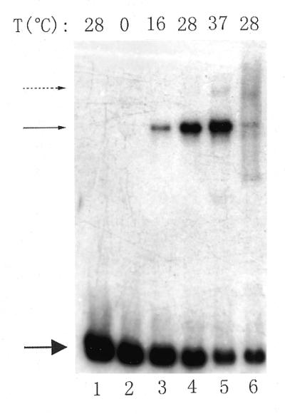
Figure 3.

Gel retardation to show that 8401 RNAP forms an open complex with the ES fragment. RNAP (1.0 µg) and labeled DNA (∼4 ng) were incubated at different temperatures, then further incubated with heparin (3.0 µg) (lanes 2–5). For lane 6, 1.0 µl of 5 mM rNTPs was added to the reaction mixture after formation of the binary complex and then was competed with heparin. Lane 1 contains the free radiolabeled DNA. The incubation temperatures are shown above each lane. The upper solid arrow indicates the RNAP–DNA open complex, the lower solid arrow indicates the free DNA and the dotted arrow denotes another heparin-resistant complex.
