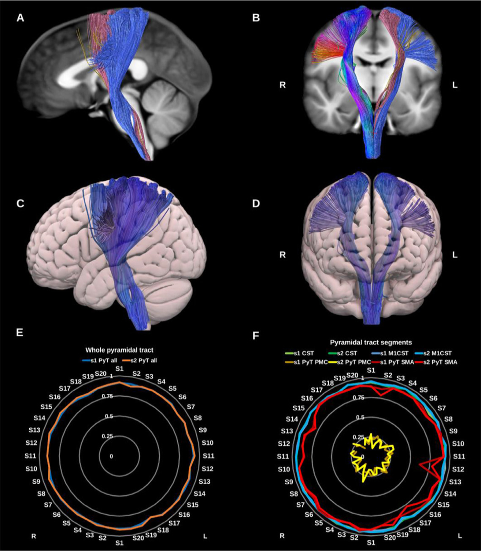
Fig. 17.

(A) and (B) whole pyramidal tract (PyT_all) overlaid in directional color coding on the right side and its different components on the left side in solid colors, premotor pyramidal tract (PyT_PMC) in yellow, supplementary motor area pyramidal tract (PyT_SMA) in pink and the corticospinal tract (CST) in blue on sagittal and coronal slices of the T1-weighted images. (C) and (D) 3D lateral and anterior projections of the semitransparent MNI pial surface with the whole PyT on both sides shown in blue. Radar plots of wDSC (vertical ranges) resulting from comparison to HCP-template bundles are shown in (E) for the whole PyT, and in (F) for the different pyramidal tract segments. L = left, R = right, S = subject, MNI = Montreal Neurological Institute, wDSC = weighted dice similarity coefficient, M1 CST = motor only CST.
