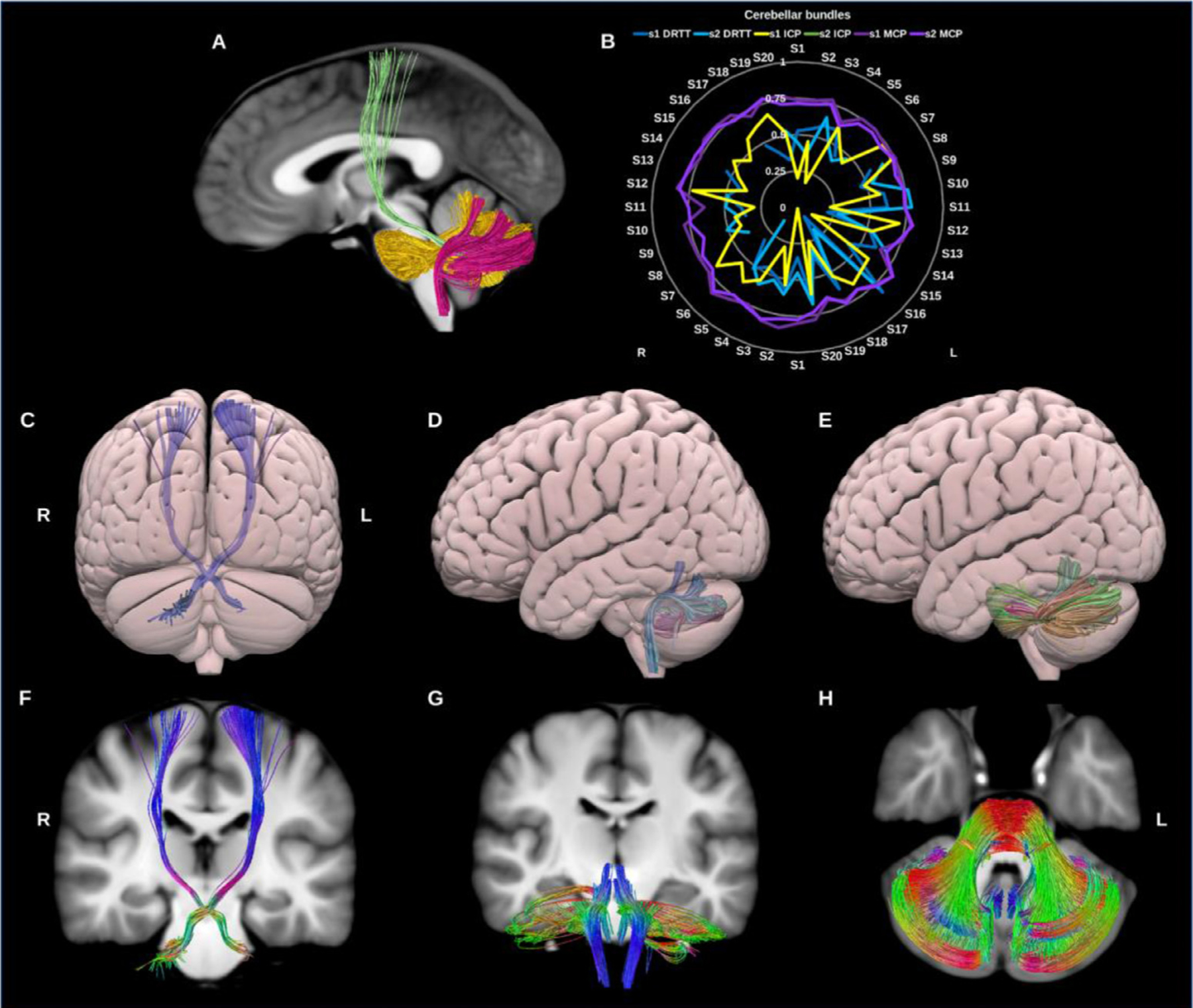
Fig. 19.

(A) Cerebellar bundles overlaid on sagittal slice of the T1-weighted images. The dentato-rubro-thalamic tract (DRTT) in green, inferior cerebellar peduncle (ICP) in fuschia, and in gold the middle cerebellar peduncle (MCP). (B) Radar plots of wDSC (vertical range) for these three bundles per session. (C), (D) & (E) show posterior and lateral surface views of the DRTT, ICP and MCP. (F), (G) & (H) show T1 coronal and axial slices of the DRTT, ICP and MCP. L = left, R = right, S = subject, wDSC = weighted dice similarity coefficient, MNI = Montreal Neurological Institute, missing results indicate failed tractography.
