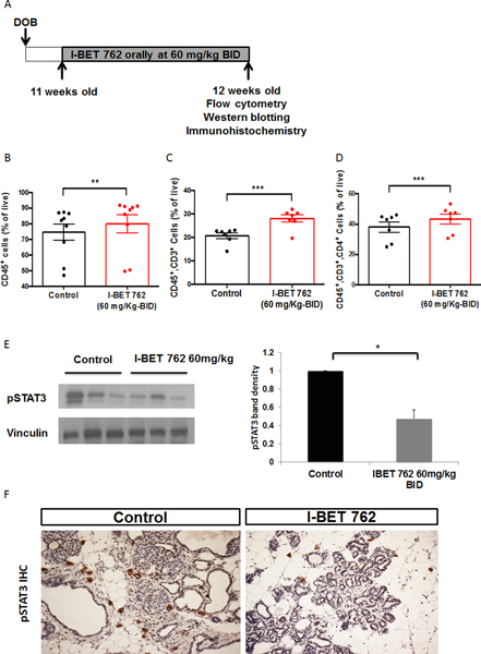
Fig 2: Short-term treatment with I-BET 762 increases CD45+ immune cells and T cell infiltration in the spleen and decreases pSTAT3 expression in the mammary gland.
A. Diagram of short-term treatment protocol by gavage (n=9 per group). The immune cell populations in mammary glands or spleen from PyMT mice were analyzed by flow cytometry. Percentages of total immune cells (CD45+), total T cells (CD45+, CD3+), and T helper cells (CD45+, CD3+, CD4+) in the spleen are shown from B to D respectively. **, p<0.01; ***, p<0.001. E. Another mammary gland from the same mice was used to isolate total proteins. Lysates were immunoblotted with anti-pSTAT3 antibodies. Representative immunoblots are shown on the left and quantification of all samples by ImageJ is shown on the right. Results were normalized to vinculin and then expressed as fold control. *, p<0.05 vs control. F. Another mammary gland in the mice was fixed in formalin and sectioned for immunohistochemistry (IHC). pSTAT3 staining is indicated in brown. Representative images are shown in F (200X magnification).

