Fig. 1.
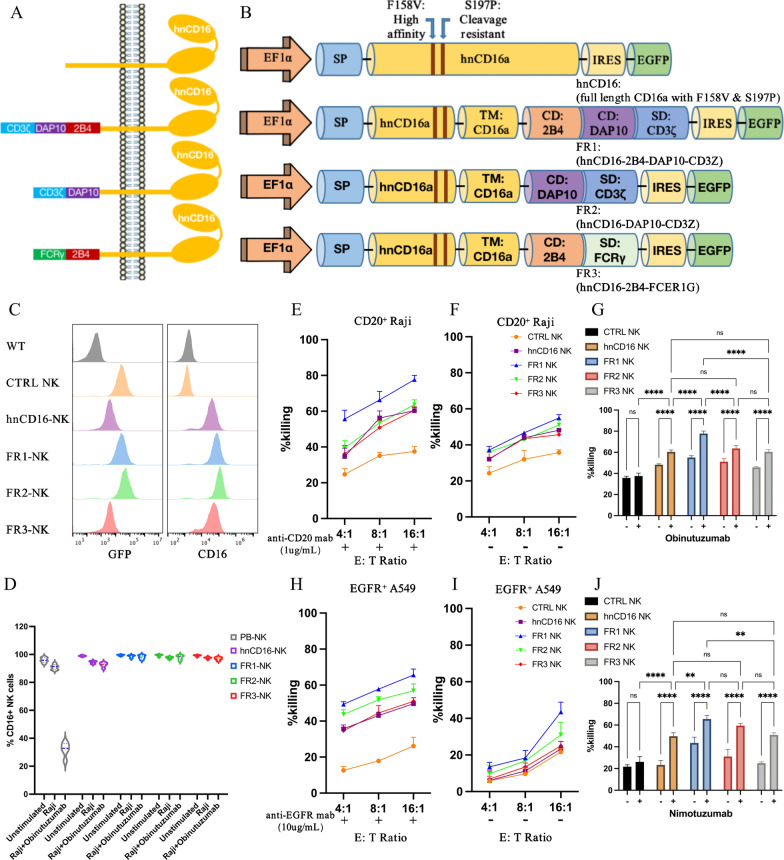
CD16-based fusion receptor constructs mediated anti-tumor activity in NK cells. A–B Schematic representation of the vector structure encoding the fusion receptor. Transmembrane (TM), co-stimulating domain (CD), stimulation domain (SD). C Expression of GFP and surface expression of CD16 in NK cells. D PB-NK cells, hnCD16-NK cells, or hnCD16FR-NK cells were stimulated for 6 h, and expression of the CD16 was determined by flow cytometry (n = 5 per group). E–G NK cells that express the designed FRs were co-cultured with Raji cells, with E or without F anti-CD20 antibody, for 12 h. ADCC was analyzed using a standard luciferase-based bioluminescence assay. The mean of percentages of specific killing ± SD are shown. G Statistical significance at 16:1 E:T ratio was determined by two-way ANOVA. Results are representative of three independent experiments. p > 0.05 (ns), p ≤ 0.05 (*), p ≤ 0.01 (**), p ≤ 0.001 (***), p ≤ 0.0001 (****). H–J NK cells that express the designed FRs were co-cultured with A549 cells, with (H) or without (I) anti-EGFR antibody, for 12 h. ADCC was analyzed using a standard luciferase-based bioluminescence assay. The mean of percentages of specific killing ± SD is shown. J Statistical significance at 16:1 E:T ratio was determined by two-way ANOVA. Results are representative of three independent experiments. p > 0.05 (ns), p ≤ 0.05 (*), p ≤ 0.01 (**), p ≤ 0.001 (***), p ≤ 0.0001 (****)

