Fig. 2.
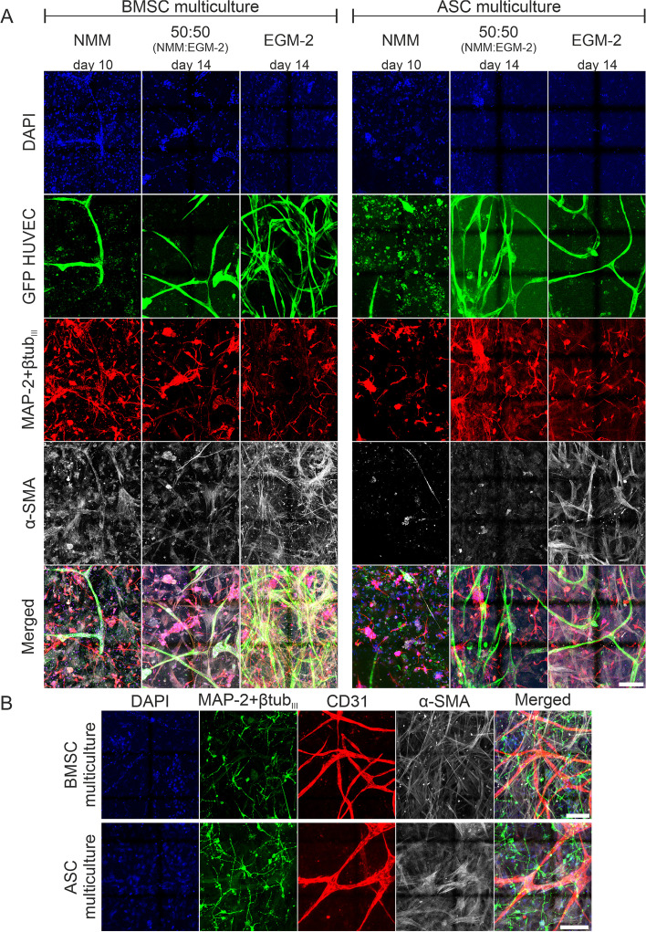
Medium optimization to support all the cell types in the multicultures. ICC staining of the BMSC and ASC multicultures on the microfluidic chip in different cell culture media on Day 10 (NMM) or Day 14 (50:50 (NMM:EGM-2) and EGM-2). a The GFP tag in the HUVECs (GFP HUVEC, green) showed that the formation of the vascular structures in both multiculture formats was best in EGM-2. The staining showed neuronal morphology (MAP-2 + βtubIII, red) in all the tested media. Pericytic properties of BMSCs and ASCs (α-SMA, gray) were greatest in EGM-2. b The staining for vascular differentiation marker (CD31, red) showed evidence of vasculature formation in EGM-2 in both multiculture formats on day 14 of culturing. Scale bar is 100 µm

