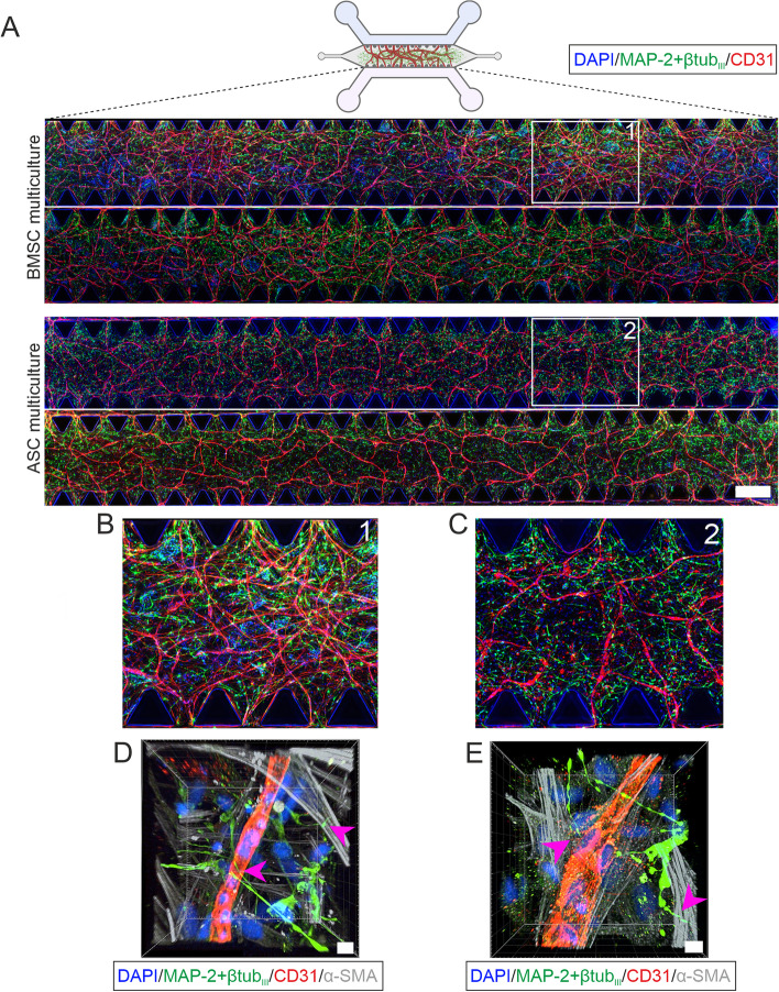
Fig. 4.
ICC staining of neurovascular networks on chip at day 14 timepoint. a Tilescan images showing the BMSC and ASC multicultures. Staining for a vascular differentiation marker (CD31, red) showed consistent and repeatable vasculature formation throughout the microfluidic chip in the BMSC and ASC multicultures. Staining for neuronal markers (MAP-2 + βtubIII, green) showed that a neuronal network had also formed throughout the hydrogel in both multiculture formats. Scale bar is 500 µm. b Close-up image of the BMSC multiculture. c Close-up image of the ASC multiculture. d-e Confocal 3D rendering of the multicultures showing neurons (MAP-2 + βtubIII, green) in the connections to both HUVECs (CD31, red) and mural cells (α-SMA, gray) in both the d BMSC and e ASC multicultures. Pink arrowheads indicate the neuronal connections to ECs and mural cells. Scale bar is 10 µm. The illustrations were created using Biorender.com

