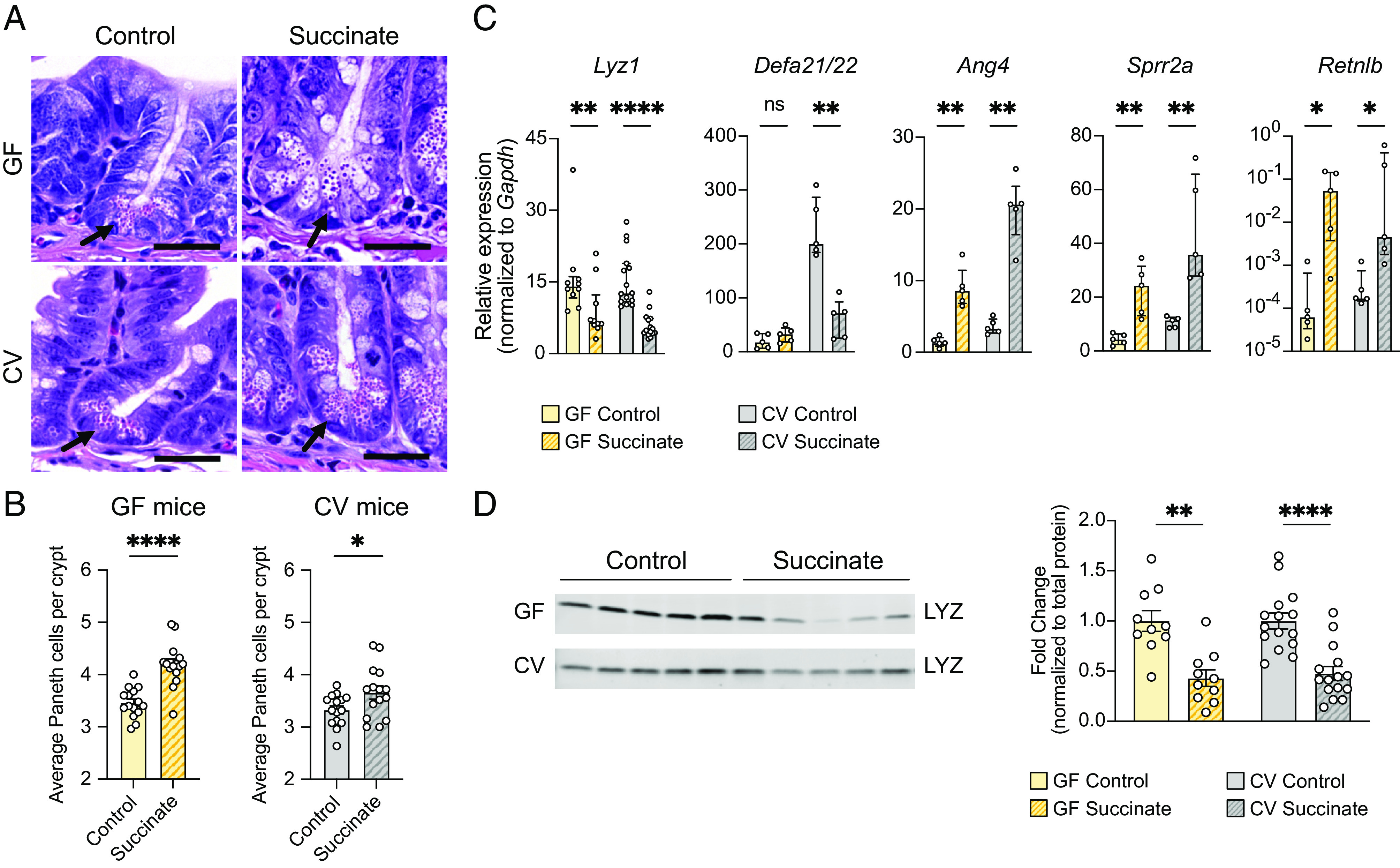
Fig. 4.

Oral administration of succinate results in similar changes to small intestinal AMP production as T. mu colonization. (A) Representative images of H&E-stained sections of the ileal crypts from control or succinate-treated GF (top row) or CV mice (bottom row). (Scale bar: 25 µm.) (B) Average number of Paneth cells per crypt in the ilea of control or succinate-treated GF or CV WT mice (n = 15 mice per group). Center values = arithmetic mean; error bars = SEM. Significance was determined using Student’s t test. (C) Expression of representative AMP genes determined by qRT-PCR in the ilea of control or succinate-treated GF and CV mice (n = 5 to 15 mice per group). Relative expression normalized to Gapdh. Center values = median; error bars = IQR. Significance was determined using the Mann–Whitney U test. (D) Representative western blot images and quantitative analysis of intracellular LYZ levels in the ilea of control or succinate-treated GF and CV mice. Each band or symbol represents an individual mouse. LYZ levels normalized to REVERT total protein stain (SI Appendix, Fig. S5F) (n = 10 to 15 mice per group). Center values = arithmetic mean; error bars = SEM. A linear mixed model was used to determine significance. ns = no significance, *P < 0.05, **P < 0.01, ****P < 0.0001.
