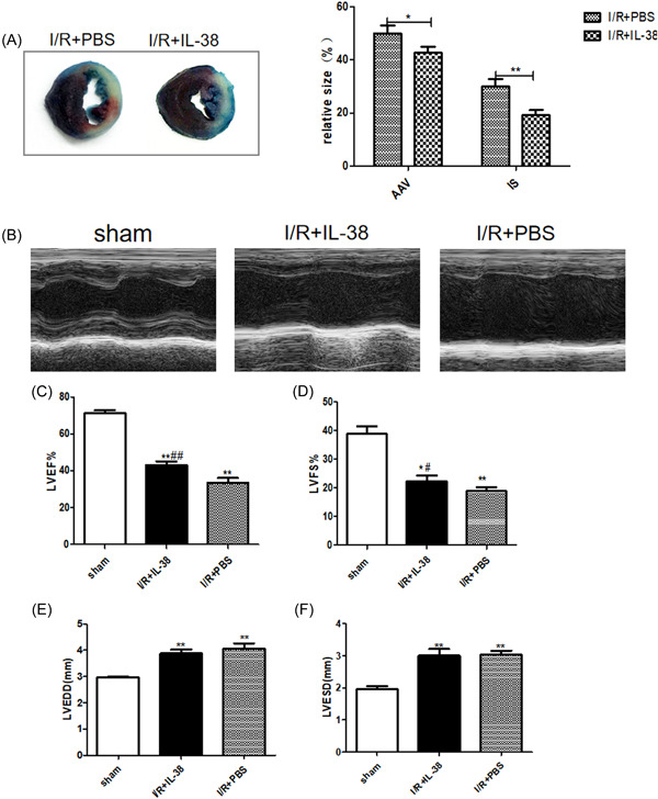
Figure 2.

Interleukin‐38 (IL‐38) improves left ventricular function after ischemia/reperfusion (I/R). (A) Representative images of Evans blue and 2, 3, 5‐triphenyl tetrazolium chloride (TTC) staining of myocardium slices and calculation of area at risk and infarct size (relative to left ventricular [LV] area) on Day 1 after I/R. (B) Representative M‐mode echocardiography images of the left ventricle on Day 1 after I/R in different groups as indicated above. (B–E) LV ejection fraction (LVEF), LV fractional shortening (LVFS), LV end‐diastolic diameter (LVEDD), and LV end‐systolic diameter (LVESD) on Day 1 after I/R (each group n = 8). *p < .05 vs. sham; **p < .01 vs. sham; # p < .05 vs. I/R + phosphate‐buffered saline [PBS]; ## p < .01 vs. I/R + PBS.
