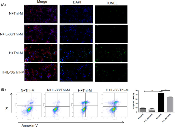
Figure 7.

Macrophages induced by interleukin‐38 (IL‐38) and troponin I (TnI) alleviated cardiomyocyte apoptosis. The supernatant of macrophages induced by TnI(TnI‐M) and IL‐38+TnI(IL‐38/TnI‐M) was used for coculture with cardiomyocytes. The mixed cells were cultured in anaerobic conditions and maintained at 37°C for 6 h and then transferred to the normoxic incubator for 6 h to undergo reoxygenation (hypoxia [H]). Cells without anaerobic conditions served as a control (normoxia [N]). (A) Representative images of terminal deoxynucleotidyl transferase dUTP nick‐end labeling (TUNEL)‐stained sections from cultured cardiomyocytes for the indicated times. TUNEL (green) and 4′,6‐diamidino‐2‐phenylindole (blue) staining of nuclei in apoptotic cardiomyocytes (red). Scar bar: 50 μm, magnification: ×400. (B). Representative images and quantitative analysis of apoptotic rate were assessed as (Annexin V (+) PI (−) cells+ Annexin V (+) PI (+) cells)/total cells × 100% using flow cytometry (n = 6). **p < .01.
