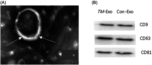
Figure 1.

Identification of exosomes derived from Talaromyces marneffei‐infected macrophages. (A) The morphology of exosomes, as shown by transmission electron microscopy. (B) Expression levels of CD9, CD63, and CD81 in macrophage‐derived exosomes stimulated by T. marneffei (TM‐Exo), as shown by western blot analysis. Con‐Exo, exosomes derived from naïve macrophages.
