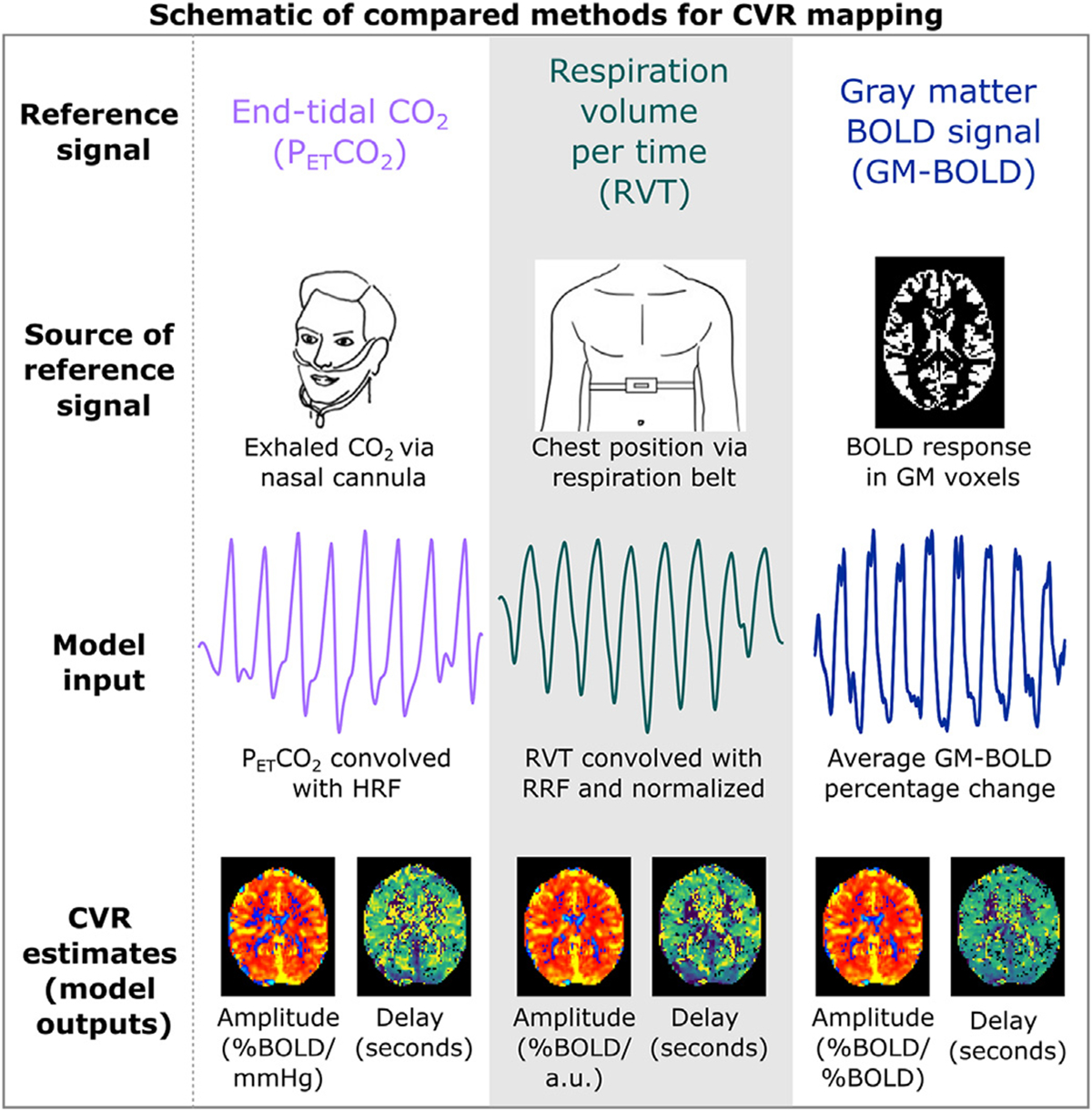
Fig. 1.

Key steps of the CVR modeling methods compared in this manuscript. Reference timeseries are generated via external recordings or the BOLD MRI data. PETCO2 and RVT timeseries are convolved with canonical response functions. For all methods, modeling is repeated for shifted variations of each reference time signal. On a voxelwise basis, the shift that optimizes the full model R2 is selected. Maps of amplitude and delay are then generated using these parameters. PETCO2 = partial pressure of end-tidal CO2, RVT = respiration volume per time, BOLD = blood oxygenation level dependent, GM = gray matter, HRF = hemodynamic response function, RRF = respiration response function.
