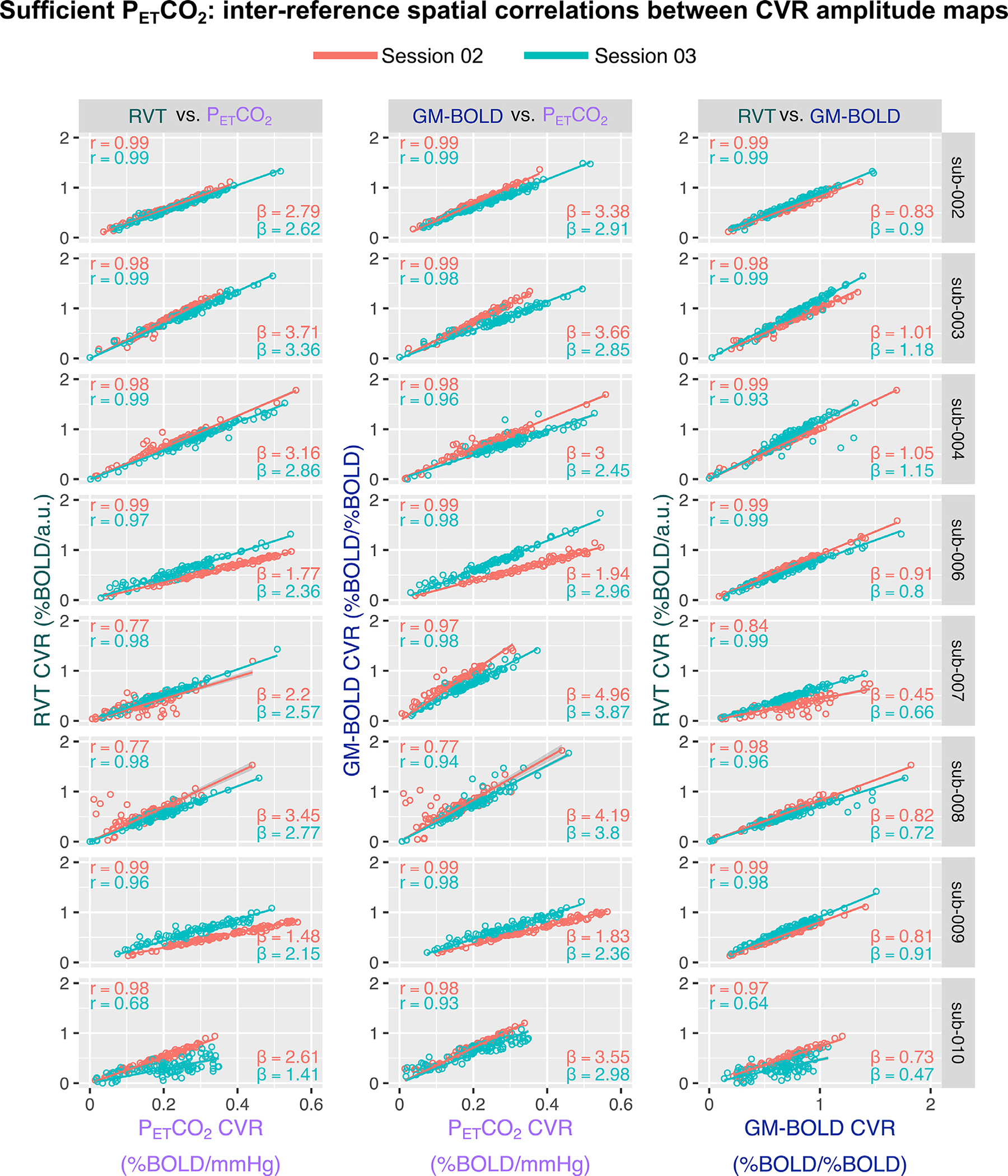
Fig. 5.

Inter-reference spatial correlations between PETCO2, RVT, and GM-BOLD CVR amplitude maps, for each subject and session (summarized in Supplementary Table S4). Each of the three pairwise comparisons are plotted in a different column. All correlations were computed using the median CVR amplitude in 96 cortical parcels, identified from the Harvard-Oxford cortical atlas and separated by hemisphere. Each dot in a sub-plot represents the median CVR amplitude in one cortical parcel. Lines-of-best-fit are shown between each pair of CVR amplitude maps. Pearson correlation coefficients (r) are listed in the top left corner and slopes for the lines-of-best-fit (β) are displayed in the bottom right corner.
