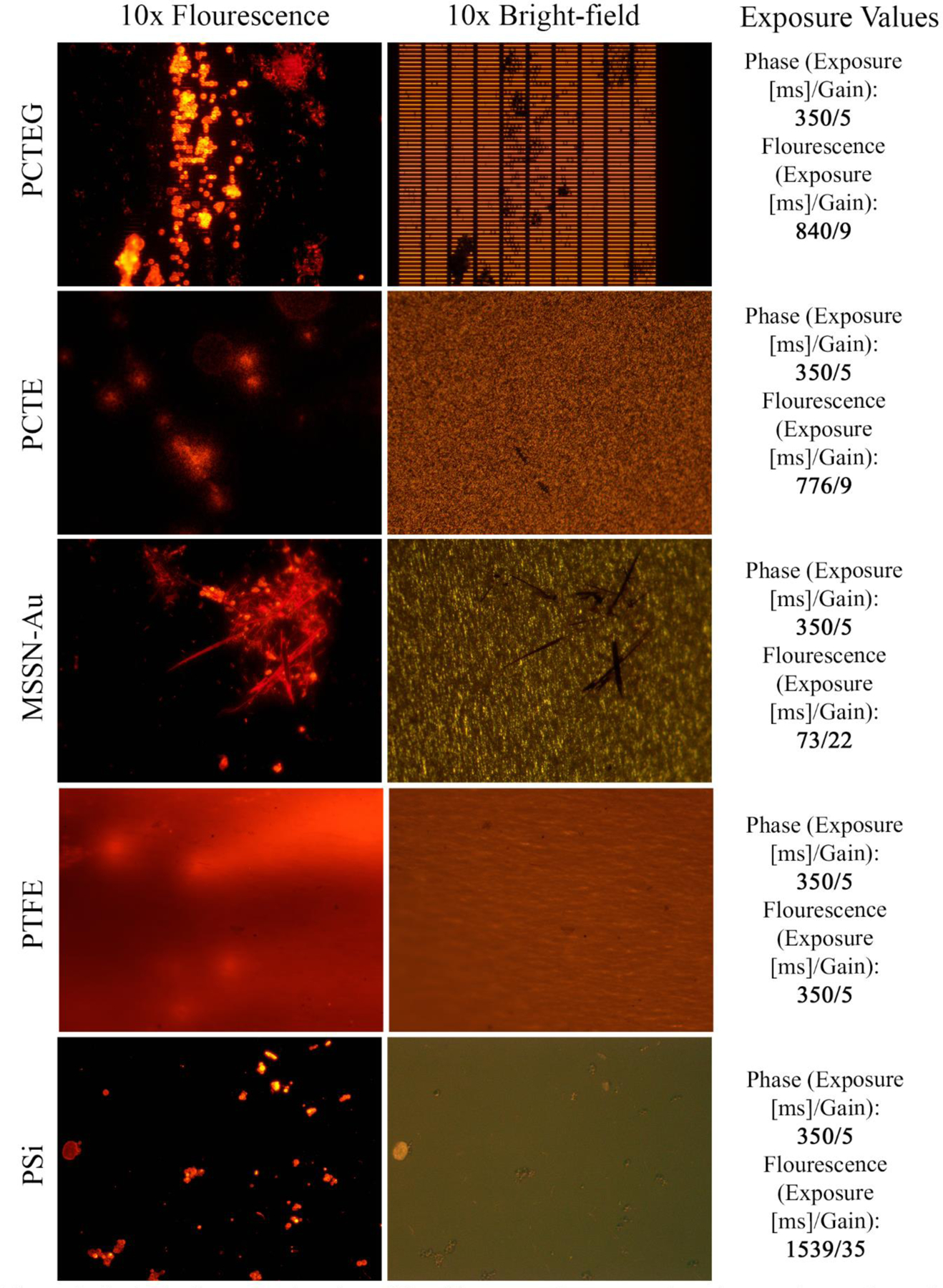
Figure 2:

Optical microscopy imaging of five filter types. Representative micrographs are shown for fluorescence (left), fluorescence-illuminated bright-field (middle), and related image acquisition data (right). Exposure time and camera gain settings are reported for each fluorescent image.
