
Fig. 6.
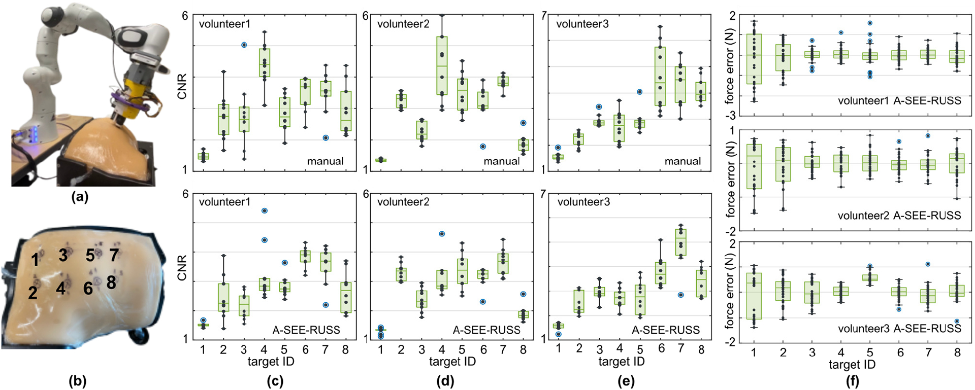
US image quality and contact force control user study. a) Experiment setup where the robot scans the LUS phantom. b) The eight pre-identified imaging targets on the lung phantom, labeled by black crosses for the study volunteers’ reference. c-e) The image quality (CNR) of the US images collected from eight targets (ten images from each target) using freehand scanning (top) and A-SEE-RUSS (bottom) by volunteer1 (professional sonographer), volunteer 2 (inexperienced operator), and volunteer 3 (inexperienced operator) respectively. f) The recorded force control error when scanning each of the targets under the phantom’s respiratory motion for the three volunteers.