
Fig. 7.
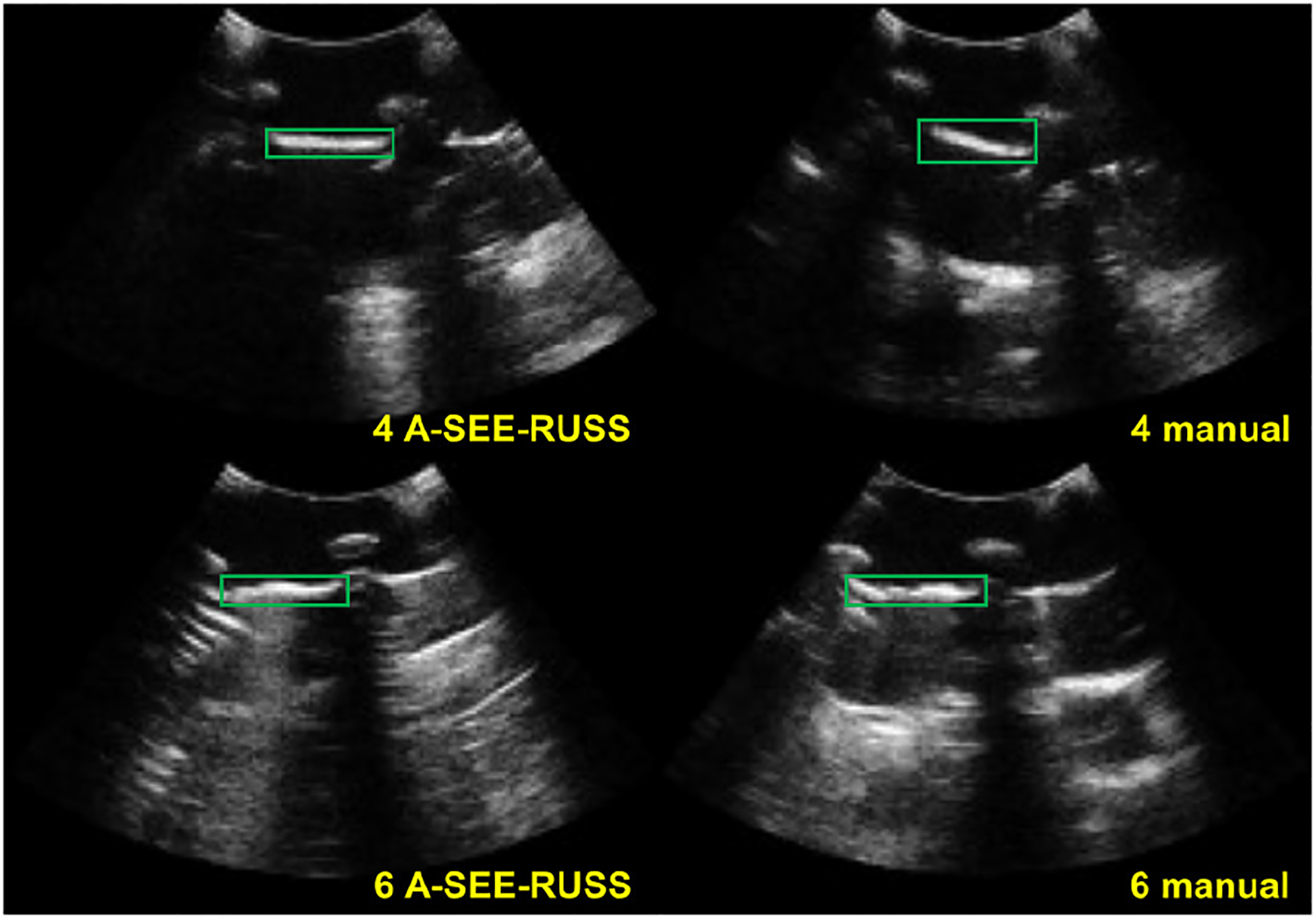
Representative US images from imaging target 4 and 6 acquired by volunteer 1 (professional sonographer) using freehand scanning and A-SEE-RUSS. At target 4, the CNR of the pleural line (bounded by green rectangles) in the manually collected image is higher than the one collected by A-SEE-RUSS. At target 6, it is the opposite.