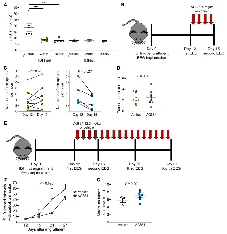
Figure 6. IDHmut inhibitors reduce seizures in IDHmut glioma–engrafted mice.
(A) IDHmut or IDHwt mouse glioma was treated with vehicle or 30 or 100 nM AG881 for 2 days in vitro. Cell pellets were collected, and intracellular levels of 2HG were assessed with mass spectrometry and normalized to total protein of each cell pellet. IDHmut glioma, n = 4 biological replicates; IDHwt glioma, n = 2 biological replicates. Bars represent mean ± SEM. **P < 0.01 by unpaired, 2-tailed t test (Bonferroni-adjusted P = 0.02). (B) Schematic of therapeutic study of C57BL/6 mice engrafted with IDHmut glioma and treated daily with 5 mg/kg of AG881 or vehicle. (C) EEGs of mice engrafted with IDHmut glioma and treated with vehicle (n = 8) or 5 mg/kg AG881 (n = 5) were blinded to treatment status and time point, and number of epileptiform spikes per hour was counted at baseline and at day 15. Data points represent each mouse, and lines connect the paired EEGs for a given mouse from different time points. Data were analyzed with paired, 2-tailed t test. (D) Maximal tumor diameter was measured from H&E-stained brains engrafted with IDHmut glioma treated with vehicle (n = 8) or 5 mg/kg AG881 (n = 5). Bars represent mean ± SEM. Data were analyzed with unpaired, 2-tailed t test. (E) Schematic of therapeutic study of C57BL/6 mice engrafted with IDHmut glioma and treated daily with 12.3 mg/kg of AG881 or vehicle. (F) EEGs of mice engrafted with IDHmut glioma and treated with vehicle (n = 3) or 12.3 mg/kg AG881 (n = 7) were blinded to treatment status and time point, and scored for percentage of 10-second epochs that contain epileptiform spikes. Data points represent mean ± SEM. Data were analyzed with 2-way ANOVA. (G) Maximal tumor diameter was measured from H&E-stained brains engrafted with IDHmut glioma treated with vehicle (n = 3) or 12.3 mg/kg AG881 (n = 7). Data were analyzed with unpaired, 2-tailed t test.

