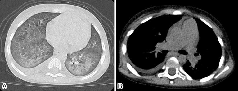
Figure 1. Chest CT findings at the onset of PVOD.
Chest CT showed infiltration, ground glass opacity, and septal thickening in the bilateral lung field along with right pleural effusion (A) and dilation of pulmonary arteries (B). No mediastinal lymphadenopathy was observed. CT, computed tomography; PVOD, pulmonary veno-occlusive disease.

