Fig. 2.

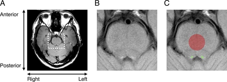
Individual examples of LC position. A) The whole image of a single participant measured by neuromelanin sensitive-weighted MRI. B) Enlarged view of brainstem area from (A). C) VOI of LC (green) and PT (red) superimposed on (B).

Individual examples of LC position. A) The whole image of a single participant measured by neuromelanin sensitive-weighted MRI. B) Enlarged view of brainstem area from (A). C) VOI of LC (green) and PT (red) superimposed on (B).