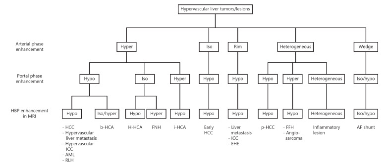
Fig. 1.
Diagnostic flowchart for HCC and its hypervascular mimics. The procedure is based on the intensity and heterogeneity of enhancement in the arterial and portal phases on CT/MRI and HBP on MRI. AML, angiomyolipoma; AP shunt, arterioportal shunt; EHE, epithelioid hemangioendothelioma; FFH, flash filling hemangioma; FNH, focal nodular hyperplasia; HBP, hepatobiliary phase; HCA, hepatocellular adenoma; b-HCA, β-catenin-activated HCA; H-HCA, HNF-1α-inactivated HCA; i-HCA, inflammatory HCA; HCC, hepatocellular carcinoma; p-HCC, poorly differentiated HCC; ICC, intrahepatic cholangiocarcinoma.

