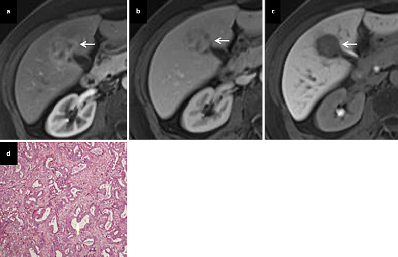
Fig. 3.
Hypervascular ICC. On dynamic contrast-enhanced MRI, a tumor (arrow) shows heterogeneous enhancement in the arterial phase (a), heterogeneous iso- and hypo-enhancement in the portal phase (b), and marked hypo-intensity in the HBP (c). A pathological examination revealed moderately differentiated ICC after hepatic resection (d).

