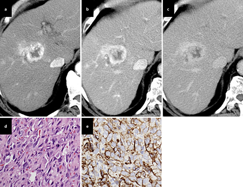
Fig. 5.
Hepatic angiosarcoma. On contrast administration, early arterial peripheral vascularity in segment V (a), a tumor was seen with centripetal enhancement in the portal (b) and equilibrium phases (c). The tumor cells are spindle-shaped, with ill-defined borders, a slightly eosinophilic cytoplasm, hyperchromatic elongated nuclei; mitotic figures are frequently seen on a hematoxylin eosin staining image (d), and CD34 immunostaining highlights the tumor cells along with blood vessels (e).

