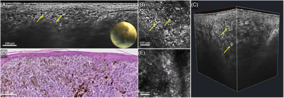
FIGURE 2.

Nodular melanoma on the lateral right upper arm of a 62‐year‐old woman: line‐field confocal optical coherence tomography (LC‐OCT) (A–C), histopathology (D) and reflectance confocal microscopy (RCM) (E). Vertical LC‐OCT examination of the pigmented area (A) showed pleomorphic melanocytes (yellow arrows) arranged in dermal nests, corresponding to the nests on histopathology (D). Horizontal LC‐OCT examination (B) showed singularly arranged atypical melanocytes (yellow arrows) under the dermal epidermal junction, which correlated well to RCM (E).
