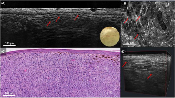
FIGURE 3.

Nodular melanoma on the lateral right upper arm of a 62‐year‐old woman: line‐field confocal optical coherence tomography (LC‐OCT) (A, B, D) and histopathology (C). Vertical, horizontal, and 3D LC‐OCT examination of the non‐pigmented area (A, B, D), revealing a hyper‐reflective and irregular wave‐like pattern with hypo‐reflective melanocytes (red arrows), which correlated well with histopathology (C).
