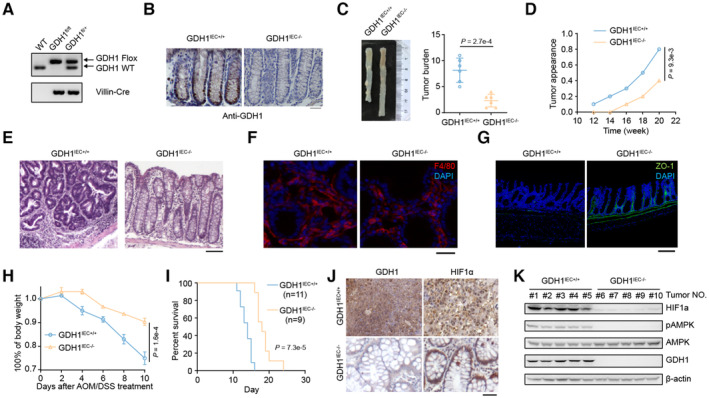
Figure 2. Depletion of GDH1 in the mouse intestine impairs colorectal cancer formation.

-
AIdentification of GDH1 knockout (KO) in mouse intestines by PCR.
-
BIdentification of GDH1 KO in mouse intestines by immunohistochemistry (IHC).
-
C–EThe AOM/DSS‐induced inflammatory cancer switch was reduced by the loss of GDH1. (C) Tumor burden (n = 6). (D) Tumor appearance. (E) H&E staining of colon cancer tumors from GDH1IEC+/+ and GDH1IEC−/− mice (scale bar, 200 μm).
-
FF4/80 staining in a noninflamed portion of the colon after AMO/DSS treatment (scale bar, 50 μm).
-
GZO‐1 staining in colon sections of mice treated with sequential AMO/DSS treatment (scale bar, 200 μm).
-
HBody weight of GDH1IEC+/+ and GDH1IEC−/− mice after AOM/DSS treatment (n = 3).
-
ISurvival percentages of GDH1IEC+/+ mice (n = 11) and GDH1IEC−/− mice (n = 9) during progression of the inflammatory cancer switch.
-
JGDH1 and HIF1α protein expression in CRC tissue from GDH1IEC+/+ mice and GDH1IEC−/− mice.
-
KBoth HIF1α protein levels and AMPK activity were assessed in the intestine of GDH1IEC−/− mice after AOM/DSS treatment using the indicated antibodies.
Data information: Data are mean ± SD from the biological replicates (C, H). Statistics: unpaired two‐tailed Student's t‐test (C); two‐way ANOVA with Tukey's HSD post hoc test (D, H); log‐rank test (I).
Source data are available online for this figure.
