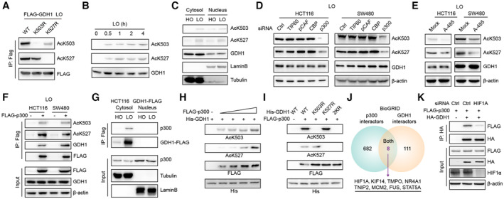
Figure 6. p300 acetylates GDH1 at K503 and K527 under hypoxia.

-
AImmunoblot analysis identified the specificity of the antibody against GDH1‐K503 or GDH1‐K527 acetylation.
-
BGDH1‐K503 or GDH1‐K527 acetylation increased with the time course of hypoxic stress.
-
CGDH1‐K503 or GDH1‐K527 acetylation occurred in both the cytosol and nucleus.
-
D, EThe levels of both AcK503 and AcK527 were analyzed when cells were transfected with small interfering RNA (siRNA) targeting TIP60, PCAF, CBP or p300 (D) or when HCT116 or SW480 cells were treated with the p300 inhibitor A‐485 (1 μM) for 12 h under hypoxia (E).
-
FHCT116 and SW480 cells were transfected with or without FLAG‐p300. The p300‐associated proteins were enriched with M2‐FLAG beads and subjected to immunoblot analysis using antibodies against GDH1, GDH1‐K503 or GDH1‐K527 acetylation.
-
GA Co‐IP assay was performed to examine the interaction between GDH1 and p300 in the cytosolic and nuclear fractions of HCT116 GDH1‐FLAG cells.
-
Hp300 acetylated GDH1 at K503 and K527 in a dose‐dependent manner.
-
Ip300 specifically acetylates GDH1 at K503 and K527.
-
JThe GDH1 and p300 interacting proteins were extracted from the BioGrid database, and the shared interacting proteins are shown in a Venn diagram.
-
KThe HIF1A gene was transiently knocked down, and HCT116 cells were cotransfected with FLAG‐p300 and HA‐GDH1 for a Co‐IP assay.
Source data are available online for this figure.
