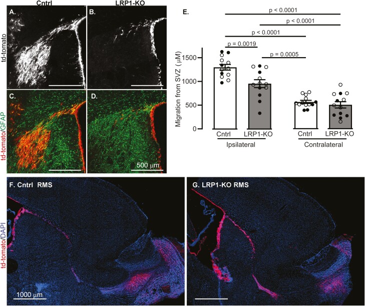
Figure 2.
LRP1-KO in adult-born neuroblasts causes migration defects after ischemia. Coronal sections show representative image of tdTomato+ cells (white) 2 weeks after MCAO in (A) control and (B) LRP1 KO mice. TdTomato images (red) were merged with GFAP-labelled imaged (green) to show post-stroke glial scarring from (C) control and (D) LRP1-KO mice. (E) Quantification of tdTomato+ cell distance from the SVZ in ipsilateral and contralateral regions (n = 13). Results are averages ± SEM. Represented significant differences were tested using ANOVA followed by Tukey’s HSD. Representative images of the RMS in sagittal sections of (F) control and (G) LRP1-KO mice.

