
Figure 1.
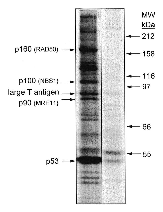
SV40 large T antigen- and p53-associated proteins in cultured SV40 large T antigen-transformed AT-1 murine cardiomyocytes. Protein complexes in NP-40 lysates from [35S]methionine metabolically-labeled cultured AT-1 cardiomyocytes were immunoprecipitated with anti-p53 monoclonal antibody PAb 421. Proteins separated on denaturing SDS–polyacrylamide gels were visualized by autoradiography. In addition to SV40 large T antigen and p53, proteins of 90, 100 and 160 kDa were consistently identified. Positions of molecular weight standards are shown in kDa on the right. The right panel is a control immunoprecipitation in which an irrelevant antibody of the same subclass as PAb 421 (IgG2a) was used in an identical manner as for PAb 421.