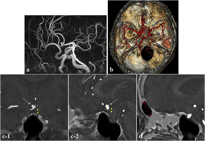
Figure 3.
Information needed to determine the surgical approach for an anterior communicating artery aneurysm (Case 2). (a) MRA showing the anterior communicating artery aneurysm and (b) Fusion image of 3D ZTE-based model and MRA showing the positional relationship of the anterior communicating artery aneurysm and bone structure of the skull base. (c-1) Coronal and (c-2) sagittal fusion images of ZTE-based image and MRA, demonstrating the height of the aneurysm from the planum sphenoidale (white arrow: aneurysm, yellow double-headed dashed arrow: distance between aneurysm and planum sphenoidale) and the size of the frontal sinus (red double-headed dashed arrow). (d) Identification of the size and location of the frontal sinus.

