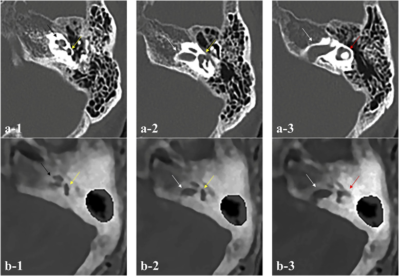
Figure 7.
Weakness of ZTE-based MRI compared with CT imaging. (a1-3) Detailed images of internal structures of pyramidal bone by CT and (b1-3) by the ZTE-based MRI in Case 5. Compared with CT imaging, exact identification of the fine structures in the inner ear including accurate location of the semicircular canal is difficult (black arrow: cochlea, yellow arrow: superior semicircular canal, white arrow: IAC, red arrow: lateral semicircular canal).

