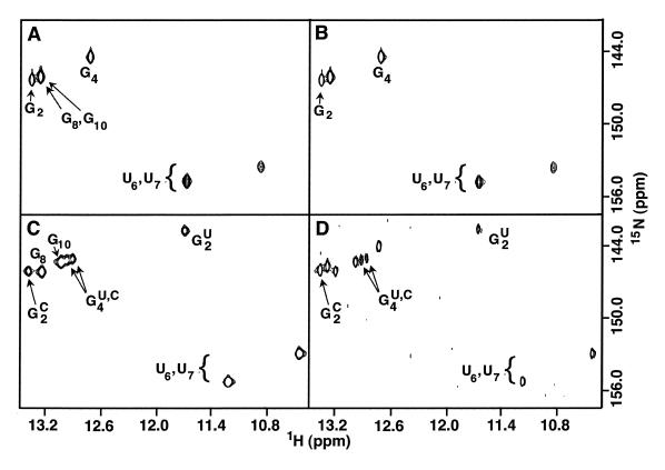
Figure 2.
Imino regions from 15N–1H HMQC spectra of (A) 15N–RNA I hairpin, (B) a mixture of 15N–RNA I and RNA IU harpins, (C) a mixture of duplexes formed by equimolar amounts of 15N–RNA I and RNA IU strands (0.40 mM each), and (D) a mixture of hairpins and duplexes formed by equimolar amounts (0.078 mM each) of 15N–RNA I and RNA IU strands. Resonance assignments for the hairpin and duplex conformations were determined using NOESY spectra. In (D), the low sample concentration and solvent exchange of the imino protons leads to very weak signals for the U6 and U7 NH resonances of the hairpin and so are not observed at this contour level. In (C) and (D), the imino resonances of G2 and G4 are labeled with C and U for the homodimer and heterodimer, respectively. The buffer conditions for (C) and (D) are identical but the 5-fold lower oligonucleotide concentration in (D) leads to partitioning between hairpin and duplex conformations.

