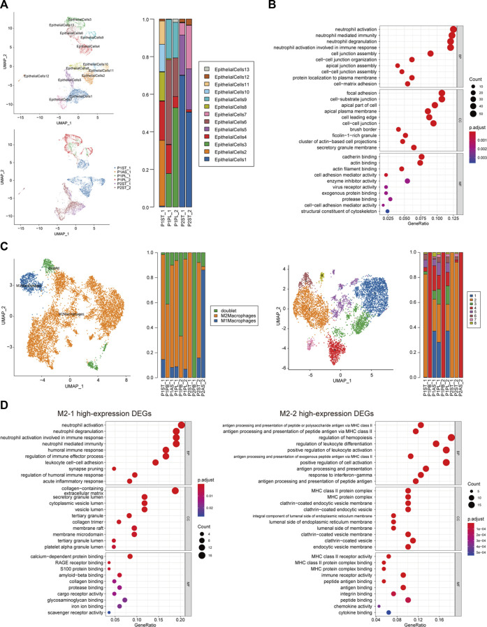
FIGURE 4.
Cellular subcluster analysis of malignant epithelial cells and M2 macrophages. (A) Cellular subcluster of malignant epithelial cells in primary lesion and pleural fluid samples; (B) dot plot of GO analysis of high-expression DEGs of malignant epithelial cell cluster 4 (EP-4); (C) cellular constitution of macrophages and subclusters of M2 macrophages; and (D) dot plots of GO analysis of high-expression DEGs of M2 macrophage cluster 1 (M2-1) and cluster 2 (M2-2).

