Figure 5.

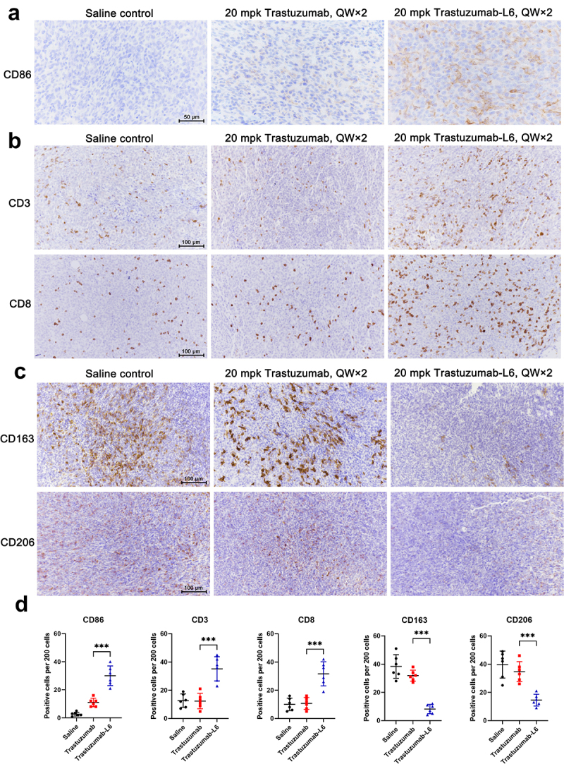
Immune cell infiltration in tumor tissue after TS-L6 treatment.
Six days after 20 mg/kg TS-L6 administration, tumor tissues were isolated, fixed, and embedded for IHC. a, A tumor from the TS-L6 treatment group exhibited more CD86+ cells compared with saline control and the TS treatment group. b, The CD3+ T cells (upper) and CD8+ T cells (bottom) infiltration was enhanced after TS-L6 treatment. c, Innate immune cells, mainly macrophages, showed decreased CD163 and (upper) CD206 (bottom) expression in TS-L6 treated tumors. D, The amounts of CD86, CD3, CD8, CD163, and CD206 positive cells were collected and the statistical differences between the TS and TS-L6-treated groups were analyzed using Student’s t test. Five independent tests were performed, and the data are shown as mean and SD.
ns, *, ** and *** represented no significance, P<0.05, 0.01, and 0.001, respectively.
