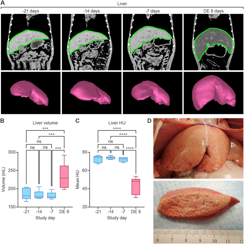
FIG 3.
Quantitative CT imaging results (rhesus monkey liver). Comparison of baseline scans and those acquired on the day of euthanasia shows hepatomegaly and decreased radiodensity after MARV exposure. (A) Coronal CT images and 3D volume renderings of the liver, based on segmentation masks. Regions of interest segmenting the liver (green borders) were automatically generated using a machine-learning-based algorithm. Data from 3 baseline imaging sessions were highly reproducible. (B) Changes in liver volume. (C) Changes in liver radiodensity (expressed in Hounsfield units [HU]). (D) Gross pathology of the liver on the day of euthanasia. The liver was markedly enlarged, with rounded edges, pale tan-yellow color, and a greasy and friable consistency (lipidosis and necrosis). ns, not significant; DE, day of euthanasia (8 days postexposure).

