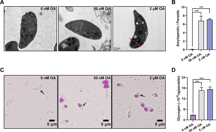
FIG 1.
Okadaic acid (OA) treatment leads to the presence of polysaccharide granules in Toxoplasma tachyzoites. (A) Transmission electron microscopy of T. gondii tachyzoites treated with 0 nM, 50 nM, or 2 μM OA for 3 h. Semicrystalline granular deposits are indicated by red arrows. (B) The numbers of polysaccharide granules observed in the tachyzoites treated with 0 nM, 50 nM, or 2 μM OA. At least 50 tachyzoites are analyzed in each group. The data are presented as the mean ± standard deviation (SD). (C) Periodic acid-Schiff staining of tachyzoites treated with 0 nM, 50 nM, or 2 μM OA. Tachyzoites are indicated by black arrows. (D) Quantification of glycogen in tachyzoites treated with 0 nM, 50 nM, or 2 μM OA. The data are presented as the mean ± standard deviation (SD). ***, P ≤ 0.001, by a t test.

