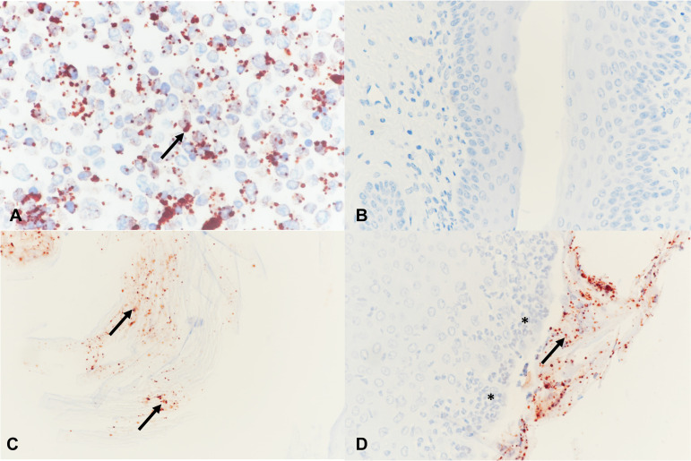
FIG 3.
Neisseria (N. gonorrhoeae) immunohistochemistry (IHC) was performed in the positive-control cell pellet (A) and in mouse vagina (B to D) with red positively labeled extracellular N. gonorrhoeae (arrows). (A) Cell pellet infected with N. gonorrhoeae. (B) Vagina negative IHC, late-sacrifice N. gonorrhoeae group. (C) Luminal N. gonorrhoeae attached to cornified epithelial cells, late-sacrifice C. muridarum+N. gonorrhoeae group. (D) N. gonorrhoeae attached to superficial layers of the vaginal epithelium, late-sacrifice N. gonorrhoeae group. The vaginal epithelium is infiltrated with PMNs (asterisks). Magnification, ×400.

