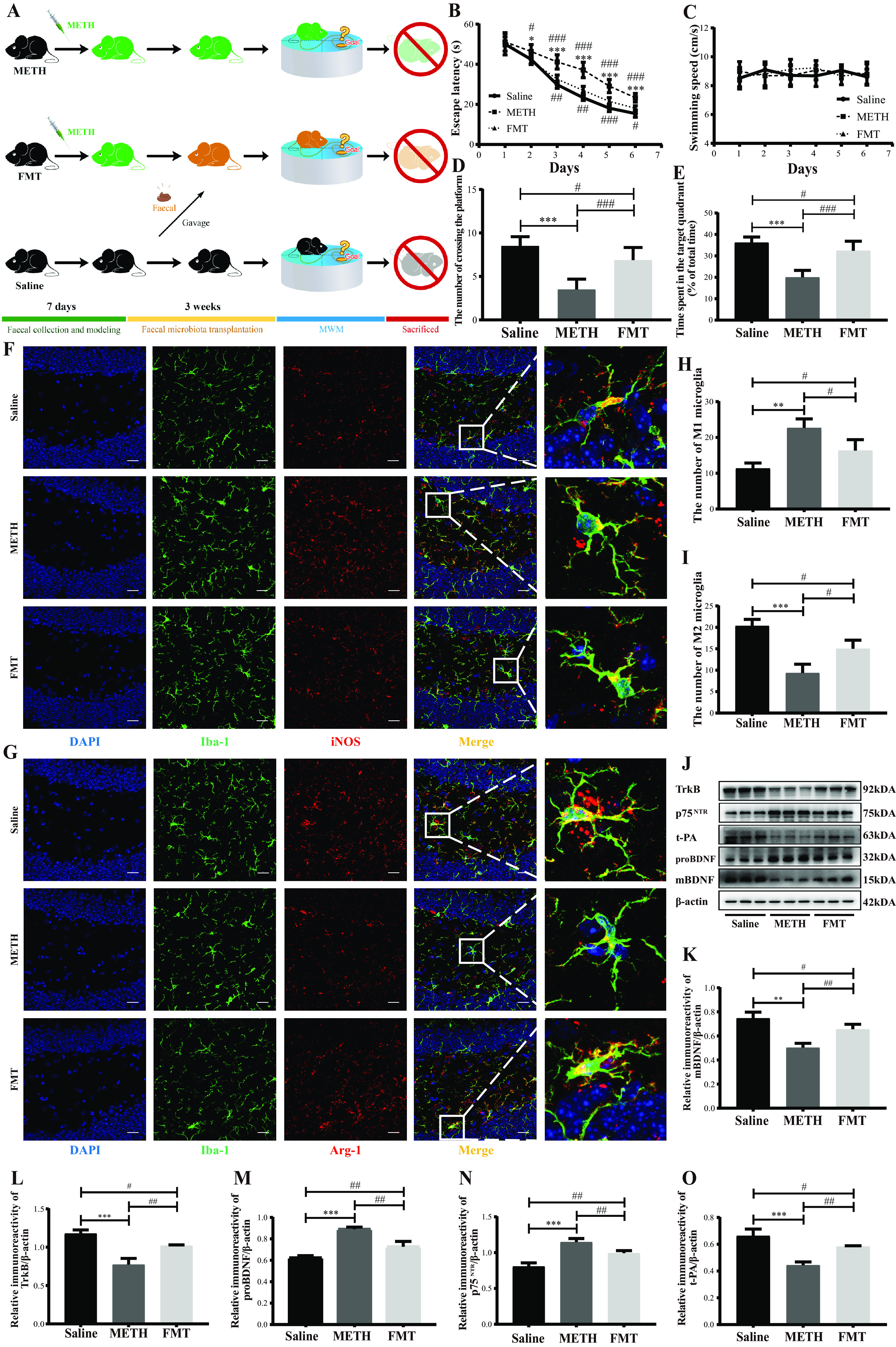
FIG 7.

Fecal microbiota transplantation attenuated chronic-METH-exposure-induced spatial learning and memory impairment by reviving microglial M1/M2 phenotype homeostasis. (A) Experimental design. Chronically METH-exposed mice were colonized with fecal samples from the mice in the saline group. (B and C) Escape latency and the swimming speed during the place navigation of the MWM test showed that spatial learning ability was attenuated by fecal microbiota transplantation from donor mice. (D and E) Numbers of instances of crossing the platform from a previously determined spot and time spent in the target quadrant (percent of total time) during the spatial probe of the MWM test indicated that spatial memory ability was attenuated by fecal microbiota transplantation from the donor mice. (F and G) Representative immunofluorescence staining images (magnification, ×400; bar = 20 μm) of nuclei (DAPI; blue), total microglia (Iba-1+; green), M1 microglia (iNOS+; red), and M2 microglia (Arg-1+; red) in the hippocampal dentate gyri of different groups. (H and I) Quantitative analysis of the number of Iba1+ iNOS+ cells (M1 microglia) and Iba1+ Arg-1+ cells (M2 microglia) in the hippocampal dentate gyri of different groups. (J) Representative protein immunoblots showing the expressions of proteins regulating the BDNF signaling system (mBDNF, TrkB, proBDNF, p75NTR, and t-PA) of different groups. (K to O) Densities of immunoblot bands of mBDNF/β-actin, TrkB/β-actin, proBDNF/β-actin, p75NTR/β-actin, and t-PA/β-actin in the hippocampi of different groups. Data are means and SEM (n = 12 to 14). *, P < 0.05, **, P < 0.01, and ***, P < 0.001, compared with the saline group; #, P < 0.05, ##, P < 0.01, and ###, P < 0.001, compared with the FMT group.
