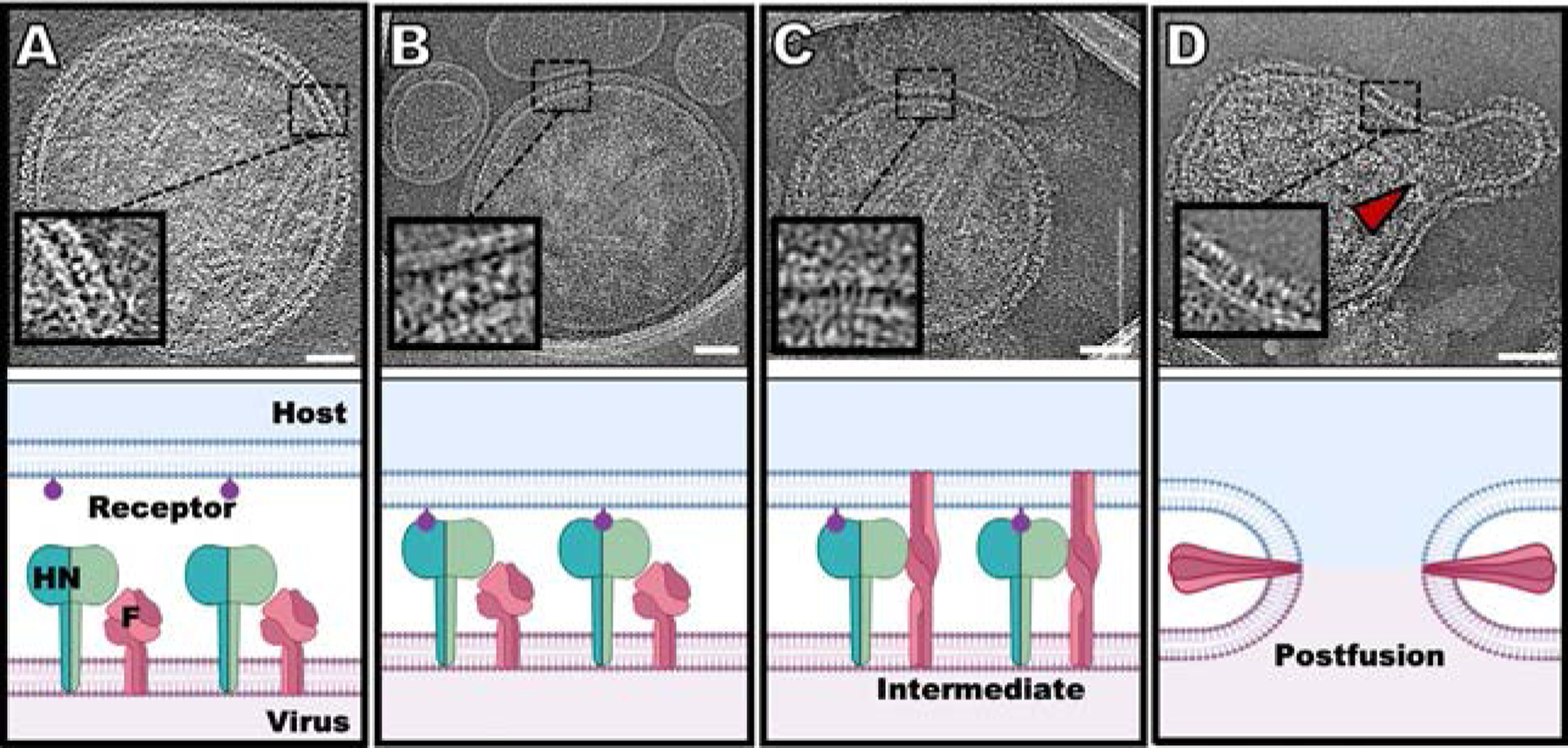
Fig. 1.

Schematic diagram of the steps in HPIV3 entry, with snapshots of cryo-electron tomographic images at each step (24). (A) HN (green) and F (dark pink) can be found densely packed on the viral surface (light pink). (B) Sialic acid (purple) binding to HN occurs in the presence of a surrogate host target membrane (erythrocyte membrane fragment) (blue). (C) Upon triggering of F by HN, F undergoes a large conformational change from a pre-fusion globular structure to a transient extended structure that crosses both membranes. (D) F folds back onto itself, pulling the viral and cell membranes toward each other, in a process that ultimately results in a merged membrane. Scale bars: (A–D) 50nm. Adapted from Marcink, T.C., Wang, T., des Georges, A., Porotto, M., Moscona A., 2020. Human parainfluenza virus fusion complex glycoproteins imaged in action on authentic viral surfaces. PLoS Pathog. 16 (9), e1008883, with permission.
