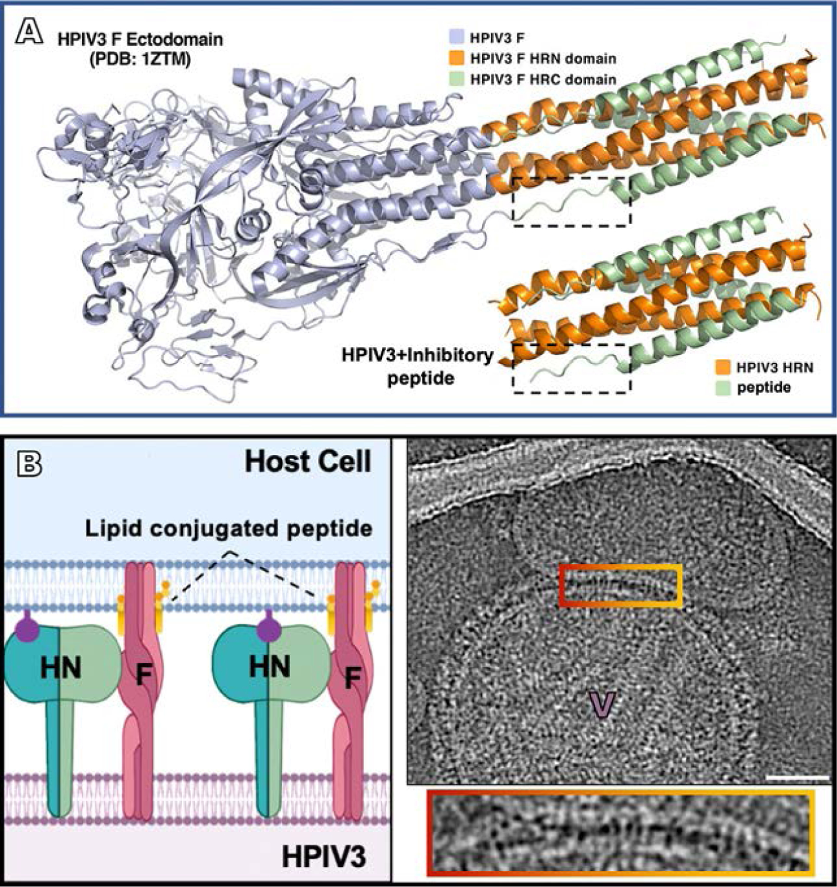
Fig. 8.

Blocking fusion at the intermediate state of F. (A) HPIV3 F ectodomain in post-fusion conformation (PDB 1ZTM), with the inhibitory peptide shown coassembled with HPIV3-HRN underneath (PDB 6NRO) (Outlaw et al., 2020a). This inhibitory peptide mimics the post-fusion state blocking F in the intermediate state. (B left panel) Schematic of lipid-conjugated inhibitory peptide inserting into the target cell membrane via their lipid tails and “locking” the extended F in its transient intermediate state, preventing refolding to the post-fusion conformation (Marcink et al., 2020b). (B right panel) Contrast-inverted images where viral particles can be observed attached to target erythrocyte fragment membranes. Enlarged region of interaction between the viral and target erythrocyte fragment membranes show elongated densities linking both membranes. Panel (A): Adapted from Outlaw, V.K., Kreitler, D.F., Stelitano, D., Porotto, M., Moscona, A., Gellman, S.H., 2020. Effects of single alpha-to-beta residue replacements on recognition of an extended segment in a viral fusion protein. ACS Infect. Dis., with permission. Panel (B): From Marcink, T.C., Wang, T., des Georges, A., Porotto, M., Moscona A., 2020. Human parainfluenza virus fusion complex glycoproteins imaged in action on authentic viral surfaces. PLoS Pathog. 16 (9), e1008883, with permission.
