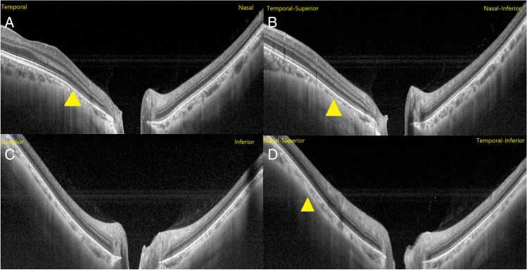
Fig. 1.
Images of PPS SS-OCT radial B-scans centered on optic disc. Image A: horizontal scan; Image B: temporal-superior to nasal-inferior scan; Image C: vertical scan; Image D: nasal-superior to temporal-inferior scan. The characteristic of an inward protrusion of the sclera compressing and thinning choroid at the staphyloma edge were observed in temporal, temporal-superior and nasal-superior (yellow triangle). The posterior sclera in the region of the PPS was bended posteriorly

