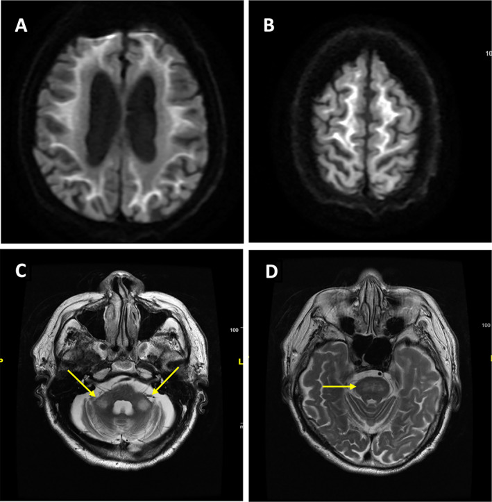
FIG. 2.

Representative brain MRI from a patient with genetically proven neuronal intranuclear inclusion disease (NIID). (A, B) Characteristic high‐intensity signal along the corticomedullary junction in the cerebral hemispheres on diffusion‐weighted imaging (DWI). High‐intensity signal on T2‐weighted images in bilateral middle cerebellar peduncles (arrows, C) and pons (D), which may mimic the radiological features of FXTAS. This was Malaysian Patient #1 in Lim et al., Ishiura et al. 101 , 102 Courtesy: Prof. Shen‐Yang Lim.
