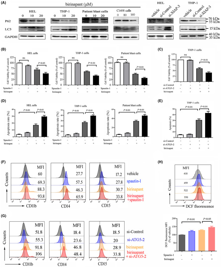
FIGURE 4.

XIAP inhibition induces autophagy as a pro‐survival signal and inhibition of autophagy enhances the anti‐leukaemic effect of XIAP inhibition in AML cells. (A) AML cell lines HEL, THP‐1 and C1498 as well as patient blast cells were treated with various concentrations of birinapant or transfected with si‐XIAP for 48 h, and then, the autophagy markers LC3‐II and SQSTM1/p62 were determined by western blot analysis. (B, D) AML cells were treated with 20 μM birinapant, either alone or in combination with 10 μM spautin‐1 for 48 h, and subsequently their cell viability was determined using CCK‐8 assay, the apoptosis was assessed by flow cytometric analysis after staining with Annexin V/PI. (C, E) THP‐1 cells were transfected with si‐ATG5‐2 or si‐Control for 24 h, and subsequently treated either alone or in combination with 20 μM birinapant for 48 h, and then the cell viability and apoptosis were determined using CCK‐8 assay and flow cytometric analysis, respectively. (F) THP‐1 cells were treated with 20 μM birinapant, either alone or in combination with 10 μM spautin‐1 for 96 h, and subsequently evaluated for the differentiation by staining with CD11b, CD14 and CD15. Images shown were representatives of at least three independent experiments. (G) THP‐1 cells were transfected with si‐ATG5‐2 or si‐Control for 24 h, and subsequently treated either alone or in combination with 20 μM birinapant for 72 h, and then, the cells were collected and evaluated for the differentiation by staining with CD11b, CD14 and CD15. (H) THP‐1 cells were treated with 20 μM birinapant, either alone or in combination with 10 μM spautin‐1 for 6 h, and subsequently ROS level was determined using DCF‐DA assay with flow cytometric analysis, the top panel is the representative images, the bottom panel is the statistical data representing three independent experiments.
